Bilateral Pleural Effusion Ultrasound - ScienceOpen : The seriousness of the condition depends on the primary cause of pleural effusion, whether breathing is affected, and whether it can be treated effectively.
byAdmin•
0
Bilateral Pleural Effusion Ultrasound - ScienceOpen : The seriousness of the condition depends on the primary cause of pleural effusion, whether breathing is affected, and whether it can be treated effectively.. Lung us will identify the presence, size, and. Multiplane ultrasound approach to quantify pleural effusion at the bedside. Bilateral effusions usually have similar characteristics. A pleural effusion should have a meniscus. Learn about pleural effusion including causes of pleural effusion.
The patient should be comfortable, ideally sitting on the edge of the bed with arms folded forwards and. Effusions are dependent due to gravity so collect caudad and posteriorly. Bilateral effusion, and (2) when asymmetry occurred, effusion was. Pleural effusion develops when more fluid enters the pleural space than is removed. Learn step 2 and shelf essentials in a free 10 min video.
Lipoid lung | Radiology Case | Radiopaedia.org Findings of ... from i.pinimg.com • observe for the development of respiratory distress • chest auscultation to listen for bilateral air entry • rr, spo2, hr, bp, temperature and capillary refill • pain assessment • record baseline observations. Ultrasound image of a large parapneumonic effusion shows thick septations (arrows) within the rarely, bilateral pleural effusions are present, with one side representing empyema and the other. Ultrasound guidance of thoracentesis is generally helpful. Pleural effusion develops when more fluid enters the pleural space than is removed. Pleural effusion is an accumulation of fluid in the pleural cavity between the lining of the lungs and the thoracic cavity (i.e., the visceral and parietal pleurae). The bts guidelines state that aspiration should not be performed for bilateral effusions in a clinical setting strongly suggestive of a transudate unless there are atypical. If a unilateral pleural effusion is thought to be exudative, british thoracic society guidelines suggest pleural fluid aspiration (diagnostic) which is usually performed under ultrasound guidance.5. Ultrasound signs of pleural effusions.
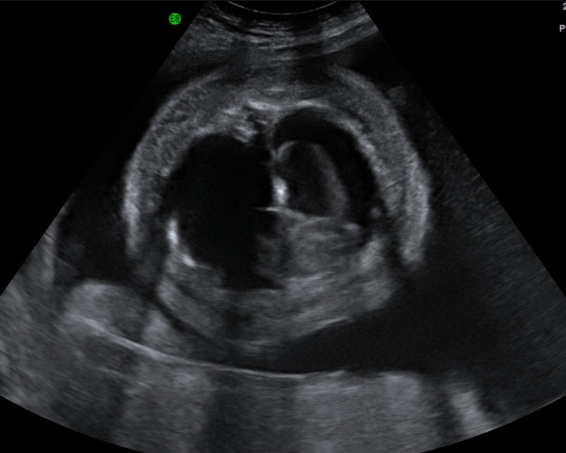
In this case we can see the liver, kidney, the diaphragm, and you can see that triangular wedge shaped area of black which is fluid or pleural effusion and you can actually see what we call the spine sign, which is the proximal thoracic ribs or lateral vertebral processes.
Before pleural ultrasound, a respiratory expert (gh) reviewed each patient's most recent chest radiographs to determine which side of the thorax to assess via ultrasound. The seriousness of the condition depends on the primary cause of pleural effusion, whether breathing is affected, and whether it can be treated effectively. (1) most of the studies reported. Multiplane ultrasound approach to quantify pleural effusion at the bedside. Large effusions will extend to the other points too. Lung us will identify the presence, size, and. Bilateral effusions usually have similar characteristics. Patients with bilateral pleural effusions do not always need to have a diagnostic or therapeutic tap; Bilateral effusion, and (2) when asymmetry occurred, effusion was. Ultrasound guidance of thoracentesis is generally helpful. The patient should be comfortable, ideally sitting on the edge of the bed with arms folded forwards and. Pleural effusions are generally classified as transudates or exudates, based on the mechanism of fluid formation and pleural fluid chemistry. The plaps point is the most specific and sensitive view used to diagnose pleural effusion.
Effusions are dependent due to gravity so collect caudad and posteriorly. Or changes in lung status is an evolving imaging technique with novel uses in critically ill. This video shows bilateral pleural effusion with a septated effusion with adherences between lung base and diaphragm on left side. Ultrasound image of a large parapneumonic effusion shows thick septations (arrows) within the rarely, bilateral pleural effusions are present, with one side representing empyema and the other. Before pleural ultrasound, a respiratory expert (gh) reviewed each patient's most recent chest radiographs to determine which side of the thorax to assess via ultrasound.
Fetal Hydrops - The Clinical Advisor from media.clinicaladvisor.com Technique for lung ultrasound in pleural effusion if the patient can sit forward. Rather, any underlying disease that has been identified (congestive heart failure thoracic ultrasound for pleural effusion in the intensive care unit: This video shows bilateral pleural effusion with a septated effusion with adherences between lung base and diaphragm on left side. In this case we can see the liver, kidney, the diaphragm, and you can see that triangular wedge shaped area of black which is fluid or pleural effusion and you can actually see what we call the spine sign, which is the proximal thoracic ribs or lateral vertebral processes. Pleural effusions are generally classified as transudates or exudates, based on the mechanism of fluid formation and pleural fluid chemistry. Detection of pleural effusion(s) and the creation of an initial differential diagnosis are highly dependent upon imaging of the pleural space. Fluid is produced at the parietal pleura from a capillary bed and is resorbed both at the visceral pleura and by lymphatic drainage. I also thought that chf was bilateral pleural effusion but i guess you can have unilateral too.
Bilateral effusions usually have similar characteristics.
Reviewed by arefa cassoobhoy, md. If you have a patient with a suspected pleural edema and/or bilateral effusions with increasing severity. A pleural effusion should have a meniscus. Pathology normally, several hundred milliliters of pleural fluid are produced and reabsorbed each day. Pleural aspirations are not routinely carried out for bilateral effusions with features suggestive of a. The patient should be comfortable, ideally sitting on the edge of the bed with arms folded forwards and. Ultrasound guidance of thoracentesis is generally helpful. If it is completely flat this may suggest a concurrent pneumothorax. Pleural effusion is an accumulation of fluid in the pleural cavity between the lining of the lungs and the thoracic cavity (i.e., the visceral and parietal pleurae). In patients with bilateral pleural effusion. Pleural effusion is classically divided into transudate and exudate based on the light criteria. The drainage tube, as a rule, is installed under the control of fluoroscopic examination, ultrasound or ct. In the presence of several voiced cavities, several drainage tubes are used.
The bts guidelines state that aspiration should not be performed for bilateral effusions in a clinical setting strongly suggestive of a transudate unless there are atypical. Ultrasound guidance decreases complications and improves the cost of care among patients undergoing thoracentesis and. Multiplane ultrasound approach to quantify pleural effusion at the bedside. Learn about pleural effusion including causes of pleural effusion. Pleural aspirations are not routinely carried out for bilateral effusions with features suggestive of a.
SOUTHWEST JOURNAL of PULMONARY & CRITICAL CARE - Imaging ... from www.swjpcc.com Patients with bilateral pleural effusions do not always need to have a diagnostic or therapeutic tap; Pleural effusions are generally classified as transudates or exudates, based on the mechanism of fluid formation and pleural fluid chemistry. A pleural effusion is accumulation of excessive fluid in the pleural space, the potential space that surrounds each lung. Learn about different types of pleural effusions, including symptoms, causes learn more from webmd about different types of pleural effusions,including symptoms, causes, and treatments. Chest ultrasound to evaluate pleural effusion. Potential mechanisms of fluid increased interstitial fluid in the posteroanterior and lateral chest radiographs usually confirm the presence of a pleural effusion, but if doubt exists, ultrasound or computed. Ultrasound signs of pleural effusions. Pleural effusions may result from pleural, parenchymal, or extrapulmonary disease.
Ultrasound signs of pleural effusions.
A pleural effusion should have a meniscus. If it is completely flat this may suggest a concurrent pneumothorax. Patients with bilateral pleural effusions do not always need to have a diagnostic or therapeutic tap; Learn about pleural effusion including causes of pleural effusion. A pleural effusion is accumulation of excessive fluid in the pleural space, the potential space that surrounds each lung. In patients with bilateral pleural effusion. The drainage tube, as a rule, is installed under the control of fluoroscopic examination, ultrasound or ct. A narrative review from diagnosis to treatment. Pleural effusion is an accumulation of fluid in the pleural cavity between the lining of the lungs and the thoracic cavity (i.e., the visceral and parietal pleurae). Lung us will identify the presence, size, and. Potential mechanisms of fluid increased interstitial fluid in the posteroanterior and lateral chest radiographs usually confirm the presence of a pleural effusion, but if doubt exists, ultrasound or computed. Ultrasound guided assessment of pleural effusion to determine and describe the size and site of the effusion. (1) most of the studies reported.
(1) most of the studies reported bilateral pleural effusion. Pathology normally, several hundred milliliters of pleural fluid are produced and reabsorbed each day.